what is cytokinesis What is cytokinesis? (with pictures)
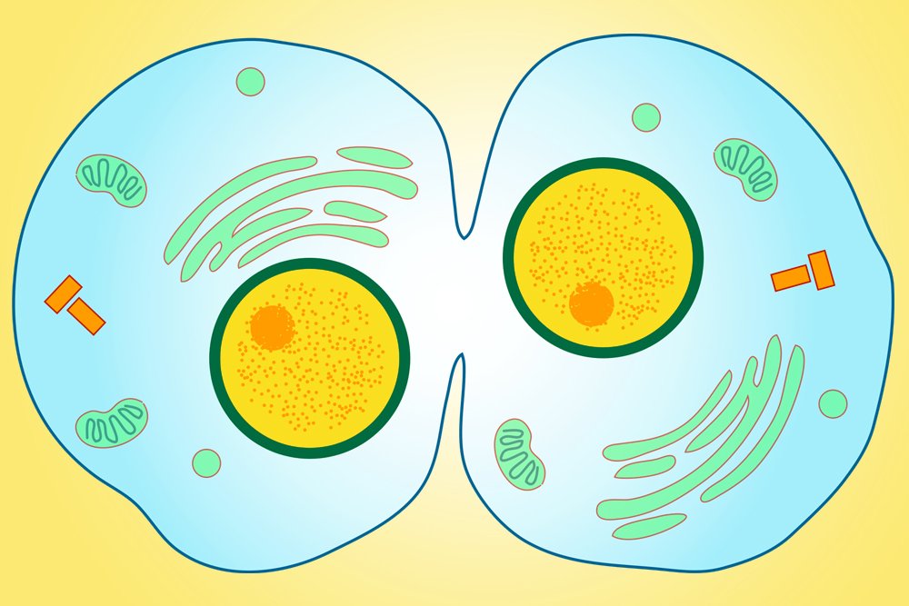
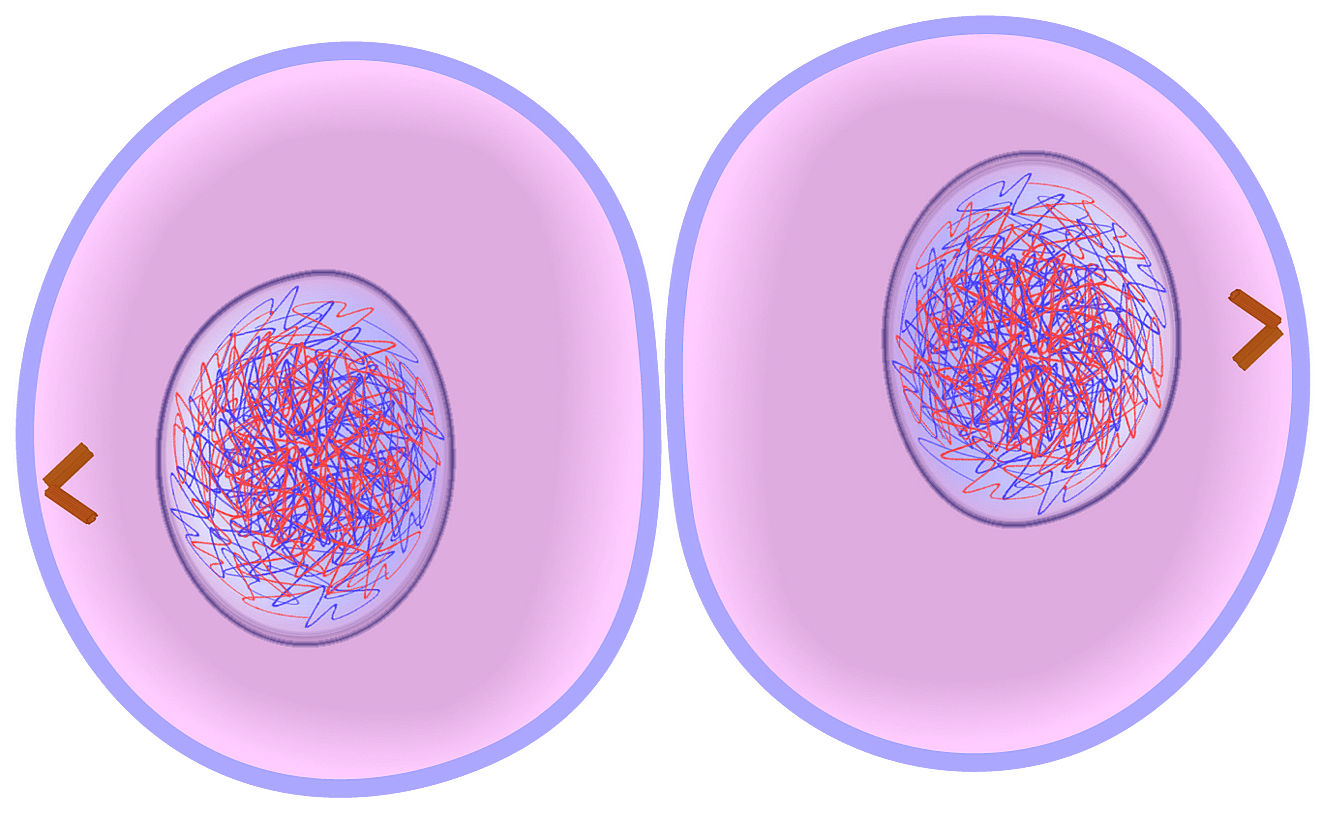
Creating new cells is an essential process for all living organisms. The division of cells into two or more daughter cells, also known as mitosis, is the process where cells replicate their DNA and split into two identical cells. However, before a new cell can be formed, it’s crucial that cytokinesis occurs. Cytokinesis is the physical separation of the daughter cells and is a vital step in the cell cycle. During cytokinesis, a new cell membrane forms around each daughter cell. This process is unique as it doesn’t require DNA replication, rather the cell membrane of the parent cell is pinched together. This process is known as cleavage and is facilitated by the action of actin and myosin, two proteins involved in cell movement. The result of cleavage is the formation of two genetically identical daughter cells, each containing a full complement of genetic material. Visualization of cytokinesis can greatly assist in understanding the process. Two images of cytokinesis are particularly useful. The first image shows a diagram of the cell undergoing cytokinesis. This image illustrates the process of cleavage and the formation of two new cells. The second image, taken with a microscope, shows a stem cell undergoing cytokinesis. This image provides a more detailed view, allowing us to see actin and myosin in action, pulling the cell membrane together to create two new cells. The diagram in Figure 1 shows the process of cytokinesis, including the formation of the cell membrane and the creation of two new daughter cells. As can be seen, once the genetic material has separated, the cell membrane pinches together to create two new cells. The new cell membranes are formed through the action of vesicles that carry lipids and proteins to the cell membrane, which then fuse and form a new bilayer. Figure 2 is an actual image taken of a stem cell undergoing cytokinesis. The image shows the same process that was illustrated in Figure 1, but in much greater detail. Here, we can see actin (in green) and myosin (in red) pulling the cell membrane together, and we can see the vesicles fusing with the cell membrane to create the new bilayer. This image allows us to appreciate the complexity and subtlety of the process of cytokinesis. In conclusion, cytokinesis is an essential process in cell division. It creates two genetically identical daughter cells that can go on to carry out important biological functions. By visualizing the process, we can gain a better understanding of how cleavage, actin, myosin, and vesicles all work together to create new cells.
If you are looking for Cytokinesis: Definition, Steps, and Significance you’ve came to the right web. We have 5 Pictures about Cytokinesis: Definition, Steps, and Significance like EduPic Cell Drawings, What is Cytokinesis? (with pictures) and also Cytokinesis: Definition, Steps, and Significance. Here you go:
Cytokinesis: Definition, Steps, And Significance
 www.scienceabc.comcytokinesis definition
What Is Cytokinesis? (with Pictures)
 www.wisegeek.comcytokinesis mitosis cell cells daughter occurs two plants tissue neoplasm soft last cleave divide forming when wisegeek
EduPic Cell Drawings
 www.edupic.netcytokinesis mitosis cell division cells edupic stage cycle two chromosomes membrane dna telophase simple complete eukaryotic daughter different visit anaphase
Cytokinesis |Genetic Engineering Info
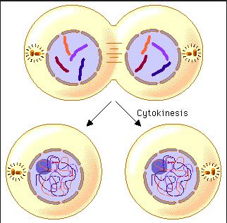 geneticengineeringinfo.blogspot.comcytokinesis telophase cell process begins
What Is Cytokinesis? (with Pictures)
 www.wisegeek.comcytokinesis mitosis facts mitogen wisegeek process cells chromatid chromatids occurs cell animal stratum following part replicate undergo plant skin
Cytokinesis mitosis cell division cells edupic stage cycle two chromosomes membrane dna telophase simple complete eukaryotic daughter different visit anaphase. What is cytokinesis? (with pictures). Cytokinesis mitosis facts mitogen wisegeek process cells chromatid chromatids occurs cell animal stratum following part replicate undergo plant skin